

In 2014, a world-renowned scientist by the name of Michio Kaku proclaimed “We’ve have learned more about the thinking brain in the last 10-15 years than in all of human history.” In the 2.5 years since Kaku wrote his piece in The Wall Street Journal, additional leaps have been made in the field of neuroscience. One such example is a recent study undertaken at Massachusetts Institute of Technology (MIT).
“Nobody has really measured neurotransmitter behavior at this spatial scale and timescale. Having a tool like this will allow us to explore potentially any neurotransmitter-related disease.” – Michael Cima, senior author of the study
In this article, we’ll delve into the extraordinary research currently underway at MIT – and how this research may indicate the next evolutionary step in the field of neuroscience.
Explaining dopamine

Dopamine is a chemical messenger (i.e. neurotransmitter) within the brain. Classified an excitatory transmitter, dopamine regulates the brain’s “reward and pleasure centers.” Here are some of dopamine’s functions in humans:
- Maintaining focus on cognitive tasks.
- Responsible for our drive and desire to accomplish tasks – or motivation.
- Enables us to see rewards and take necessary action.
Individuals diagnosed with Attention Deficit Disorder (ADD) or Attention Deficit Hyperactivity Disorder (ADHD) are often prescribed drugs that elevate dopamine levels; hence, “rewiring” the brain to focus.
Dopamine deficiencies increase the risk of Parkinson’s Disease – a degenerative disorder that affects control of the motor system. Low dopamine levels also makes one more susceptible to addiction.
The study’s abstract concisely explains the potentially adverse consequences of a neurochemical imbalance: “Dysregulation of neurochemicals, in particular, dopamine, is epitomized in numerous debilitating disorders that impair normal movement and mood aspects of our everyday behavior.”
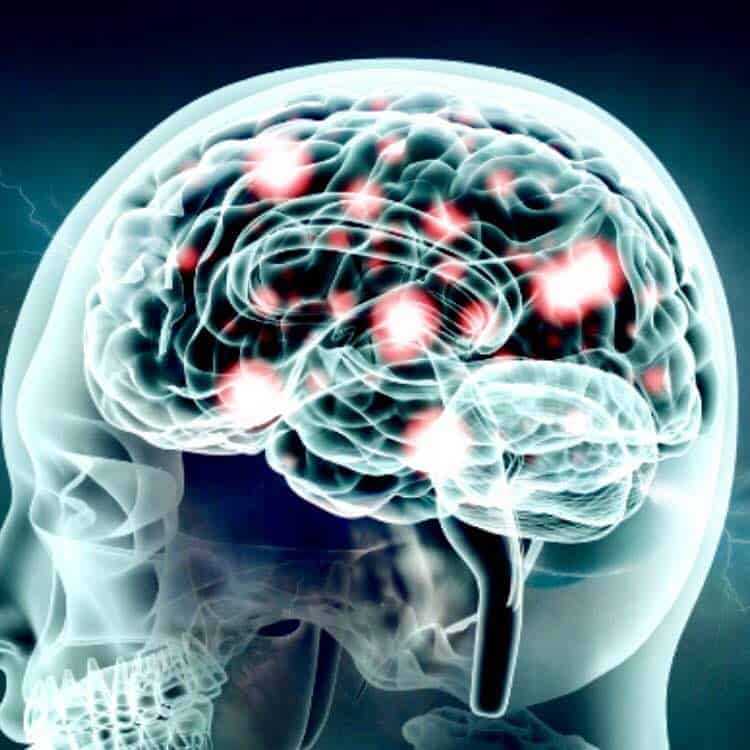
MIT Researchers Reveal Discovery That Could Evolve The Brain
Accurate measurement of neurotransmission activity within the brain is a notoriously difficult process. Neuroimaging is most commonly practiced using positron emission tomography, or PET. The problem with using PET technology for neuroimaging is two-fold: (1) PET measures metabolic processes via blood flow – a less than ideal solution for gauging chemical activity in the brain, and (2) the cost of operating PET equipment is often exorbitant.
Thus far, neuroscientists have used implanted electrodes with limited success. Despite the formidable progress of brain sensor technology, these devices have been determined unreliable due to their short duration and scar-producing effects. Furthermore, the probability of detecting any notable measure of dopamine is only 50 percent.
Neuroscientists at MIT developed and began experimenting with a new, carbon-electrode neural sensory device in rats. Unlike sensors developed prior, the MIT-designed transmitter permits for “accurate readings for longer periods of time and covering more of the brain.” MIT’s neuroimaging sensor, comprised of miniature carbon electrodes, overcomes the inadequacies of previous sensors primarily through its diminutive size.
About the sensor
The device is approximately one-tenth the size of previous brain sensors and accurately measures dopamine levels within milliseconds. These specifications may ultimately permit for widespread usage in human patients.
The sensor, which looks very similar to a microchip, consists of the following properties:
– arrays of 8 electrodes, each measuring 10 microns in diameter
– use of rigid polymer, or PEG, to allow for an adequate duration of usage
Here’s how the sensor safely and accurately measures dopamine levels:
– scientists administer an oscillating voltage via the electrodes
– at a certain voltage level, any dopamine in the area transmits a measurable electric current via an electrochemical reaction
Research Implications
Further experimentation of the transmitters continues to take place. Thus far, the device has undergone rigorous application – and has remained in working order for up to two months.
This bleeding-edge neuroimaging research could have far-reaching effects; including new insight into dopamine’s roles in learning, memory, and emotion. Ann Graybiel, one of the study’s authors, states “We and other people have struggled with getting good long-term readings. We need to be able to find out what happens to dopamine in mouse models of brain disorders, for example, or what happens to dopamine when animals learn something.”

Further collaboration efforts to develop injectable devices, such as those that transmit drugs to areas of the brain, continues to transpire. In short, this and similar research demonstrates tangible progress in the future treatment of neurodegenerative diseases.
While treatment of neurodegenerative diseases is the primary goal; successful human application of MIT’s design may also evolve the treatment of anxiety, learning disorders, and many other adverse conditions attributed to dopamine deficiency.
Helen Schwerdt, one of the study’s authors and a fellow at MIT’s Koch Institute for Integrative Cancer Research, states “What links all these studies together is…to find a way to chemically interface with the brain. If we can…it makes our treatment or our measurement a lot more focused and selective, and we can better understand what’s going on.”